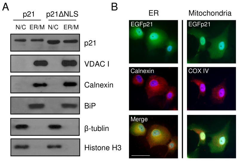
Figure 4. P21 localizes to an ER/mitochondria-enriched subcellular fraction.
H1299 cells with inducible expression of EGFp21 (p21) or EGFp21ΔNLS (p21ΔNLS) were cultured in the presence of doxycyline for 24 hrs and exposed to hyperoxia for 2 days. (A) Cellular fractionation was performed, enriching for nuclear/cytosolic (N/C) and ER/mitochondria (ER/M) fractions. Protein lysates were isolated and immunoblotted for the p21 transgene, β-tubulin, calnexin, BiP and VDAC I. (B) EGFp21 inducible cells cultured doxycycline and treated with hyperoxia for 2 days were fixed and immunostained for EGFp21 (green) and calnexin or COX IV (red) with dapi (blue) as a nuclear counterstain. Images were captured at 400X magnification and the scale bar in the lower left image is 50 μm.

