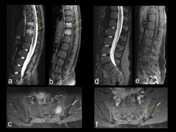
Figure 3.
Imaging data of spinal and sacral CRMO lesions in a 10-year old girl. (A-C). Pre-treatment sagital (A and B) and axial (C) MRI. (A) STIR sequence and (B and C) post-gadolinium T1-weighted sequence reveal abnormal signal in vertebral bodies of T10, T11, and S1 (arrows), as well as in sacral ala (arrow). (D-F). Post-treatment (5 months after initiation of pamidronate) MRI using the same technique as (A-C): Complete resolution of the previously seen abnormal signal.

