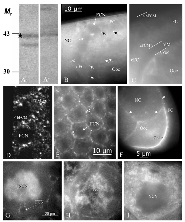
Figure 2.
Inx2 is localized to membrane plaques in follicle cells and germ-line cells and is distributed stage-specifically in nurse cells. On immunoblots of ovary extracts using Inx2-antisera (A: AInx2Rb-CT(KLRH), A': AInx2GP-CL) a band at the calculated molecular mass of 42 kDa (asterisk) and a band at 39 kDa are recognized. B: Inx2 is localized to presumed gap-junction plaques between follicle cells as well as between follicle cells and germ-line cells (stage 10b, AInx2Rb-CT(REM), optical median section, WFF; see also Fig. 4D). Black arrows point to apico-laterally situated plaques between follicle cells, white arrows point to plaques between follicle cells and oocyte, and arrowheads point to plaques between centripetally migrating follicle cells and nurse cells. C: Control follicle (NIS) showing the anterior-dorsal region of the oocyte and the follicular epithelium (comparable to the region shown in B). Different membrane regions are marked with white lines. Apical follicle-cell membranes make contact with the oolemma via microvilli spanning the developing vitelline membrane (VM). D, E: Inx2 is found in lateral and apical, but not in basal follicle-cell membranes (arrowheads, stage 11, Anti-Inx2GP-CL, D: LSM, E: optical tangential section, WFF). F: Inx 2 is located in the oolemma (arrowhead) as well as in nurse-cell membranes (white arrows, stage 8, Anti-Inx2GP-CL, optical median section, WFF). G-I: Stage-specific distribution of Inx2 in the nurse cells (AInx2Rb-CT(REM), WFF): During stage 10a (G), Inx2 accumulates around nurse-cell nuclei (NCN). During stage 10b (H), Inx2 becomes dispersed in the cytoplasm. During nurse-cell regression (stage 11, I), Inx2 is found in cytoplasmic clouds and in particles that become transported into the oocyte. FCN, follicle-cell nucleus (not stained); for further abbreviations, see legend to Fig. 1.

