Figure 3.
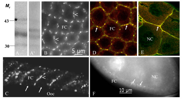
Inx3 is colocalized with Inx2 to membrane plaques in follicle cells and nurse cells. On immunoblots of ovary extracts using Inx3-antisera (A: AInx3GP-CT, A': AInx3Rb-CL) a weak band at the calculated molecular mass of 45 kDa (asterisk) and two bands at 35 and 30 kDa are recognized. B, C: Inx3 is localized to presumed gap-junction plaques between follicle cells (stage 10a). It is expressed in a continuous as well as punctate pattern at the lateral membranes (arrowheads, B: AInx3Rb-CL, WFF, C: AInx3GP-CT, LSM), but is missing at the apical membranes and at the oolemma (arrows). D, E: Inx2 (red, AInx2Rb-CT(REM)) and Inx3 (green, AInx3GP-CT) are colocalized in lateral follicle-cell membranes (yellow, arrows in D, stage 10a, LSM) as well as in nurse-cell membranes (yellow, arrow in E, stage 10b, LSM). F: In young follicles, Inx3 is located in plaques between nurse cells and follicle cells (arrows) and around nurse-cell nuclei (stage 7, AInx3Rb-CL, WFF). For further abbreviations, see legend to Fig. 1.

