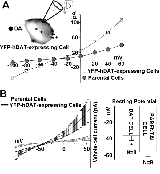
FIGURE 1.
Expression of DAT constitutively depolarizes cells. A, representative current-voltage (I(V)) relationships of DAT-mediated currents in DAT-expressing cells (open squares) compared with I(V) relationships in parental, nontransfected cells (closed circles). Cells were voltage-clamped with a whole-cell patch pipette containing 30 mm Na+ without DA present inside or outside the cells (see “Materials and Methods”). Whole-cell currents were obtained by stepping the membrane potential in -20 mV steps between -100 to +60 mV, from a holding potential of -20 mV. The I(V) curves were obtained after subtracting currents in the presence of 10 μm of the DAT inhibitor, cocaine (see “Materials and Methods”). B, left panel shows the expansion of A near the two reversal potentials. The I(V) curves were fit to polynomials (solid lines) from the reversal potentials (n = 8–9). Right panel depicts bar graphs of the mean resting potentials measured in DAT-transfected cells (-36.5 ± 5.5) and parental nontransfected cells (-56.3 ± 4.7) p < 0.05.

