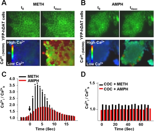
FIGURE 6.
METH causes greater increases in intracellular Ca2+ than AMPH. DAT-expressing cells exposed to METH (A) or AMPH (B) at t = 0 and 4 s. Basal fluorescence readings were taken for 60 s (∼60 images) before stimulation with either METH (10 μm) or AMPH (10 μm) as described under “Materials and Methods” ([Ca2+]b). C, internal free Ca2+ ([Ca2+]i) after adding 10 μm METH or AMPH (solid arrow) to the DAT-expressing cells; n = 97–151 cells measured in 3–5 independent experiments (0.001 < p < 0.05). D, cocaine pretreatment blocks the effect of METH and AMPH on [Ca2+]i.

