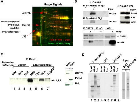
FIGURE 4.
ARF interacts with Bcl-xl. A, 2D-DIGE of mitochondrial ARF-interacting proteins in U2OS-ARF cells. The red spots indicate ARF-interacting proteins; the box depicts the sequence of three tryptic peptide fragments from Bcl-xl obtained from the mass spectrometry analysis. B, co-immunoprecipitation of ARF with antisera to Bcl-xl (H5, Santa Cruz Biotechnology, or anti-Bcl-xl, Cell Signaling) and GRP75 in U2OS-ARF cells. Doxycycline treatment (Doxy) was for 24 h. On the right, the level of Bcl-xl, ARF, and actin in whole cell lysate from the same samples is depicted. C, co-immunoprecipitation of endogenous ARF with Bcl-xl from mitochondrial (Mito), cytosolic (Cyto), or whole cell lysate (WCL) extracts from MEFs infected for 48 h with parental retrovirus (vector), or retrovirus expressing E1A and Ras (E1a/Ras) plus short hairpin to silence p53 (shp53). In the panel on the right, 20 μg of mitochondrial and cytosolic extracts were probed for the mitochondrial proteins GRP75 and Bak, and the cytosolic/nuclear protein PCNA to attest to the purity of these mitochondrial fractions. D, in vitro GST binding assay using GST or GST-ARF and the 35S-radiolabeled proteins Bcl-xl, Mdm2, or BAK. 20% of the input of radiolabeled proteins from each in vitro transcription/translation reaction is on the left (4 μl of a 50 μl transcription/translation reaction), and a Coomassie staining of 1 μg of purified GST fusion proteins is depicted on the right.

