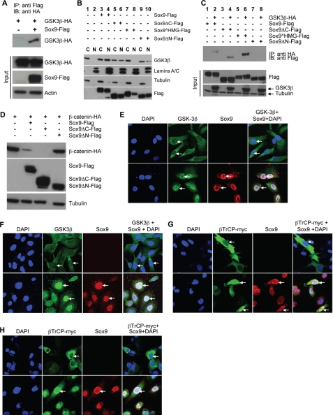
FIGURE 6.
Sox9 binds GSK3β and promotes its nuclear translocation.
A, GSK3β-HA was co-immunoprecipitated (co-IPed) with
Sox9-Flag when transiently co-expressed in CHO cells. B, CHO cells
were transiently transfected with a vector alone or the indicated plasmids.
Cells were fractionated and subcellular distribution of endogenous GSK3β
was analyzed by Western blot by anti-GSK3 antibody. Sox9 and its mutants were
detected by anti-FLAG antibody. Lamins A/C and tubulin served as markers for
nuclear and cytoplasmic fractions, respectively.
Sox9 HMG, although at a lower
level, was also detected in the nuclear fraction, possibly due to some
contamination by the cytoplasmic fraction. C, CHO cells were
transiently transfected with the indicated plasmids. GSK3 was
immunoprecipitated using anti-HA antibody. Sox9 was detected with anti-FLAG
antibody. D, CHO cells were transiently transfected with
β-CATENIN-HA and the indicated plasmids and analyzed by Western
blot. Tubulin served as loading control. E and F, NCI-H28
(E) or SNU475 (F) cells were infected with
Sox9-adenovirus and localization of endogenous GSK3β
(green) and Sox9 (red) were detected 24 h later. DAPI
stained the nucleus (blue). G and H, NCI-H28
(G) or SNU475 (H) cells were transfected with
βTrCP-myc and infected with Sox9-adenovirus when
indicated. Localization of βTrCP-myc (green) and Sox9
(red) was detected by immunofluorescence. DAPI stained the nucleus.
IB, immunoblot.

