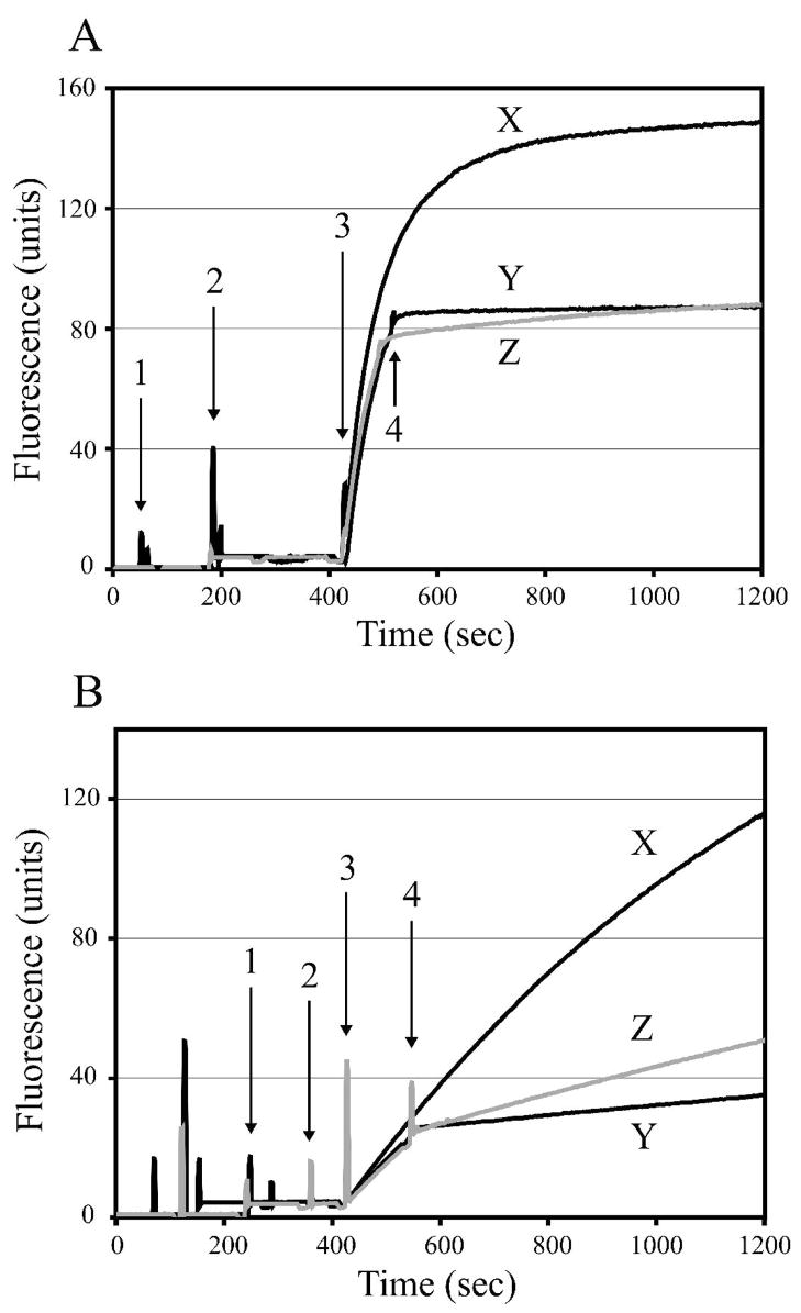
Figure 2.
Inhibitory effect of 1 and 2 on m-calpain (A) and μI–II (B). Duplicate assays were performed by adding 1.3 μM (EDANS)-EPLFAERK-(DABCYL) (1), 125 nM calpain (2) and 4 mM CaCl2 (3), then averaged to yield the plots shown. Autolytic inactivation of m-calpain, but not μI–II, was observed when no inhibitor was added (X). Immediately following addition of each inhibitor (4), the increase in fluorescence intensity was attenuated. 1(Y) caused a more distinct attenuation of fluorescence in both the m-calpain and μI–II assays, indicating that it is a more potent inhibitor when compared to 2 (Z).

