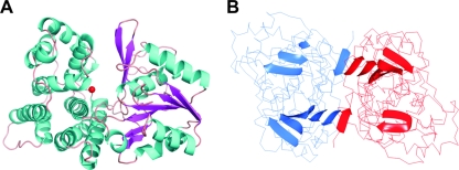
FIG. 2.
Overall structure of the monomer and dimer of 1,3-PD dehydrogenase. (A) Ribbon representation of the enzyme monomer colored by secondary structure elements; the Fe atom is represented by a red ball. (B) Main-chain trace of the dimer structure with the location of the beta strands in each monomer. The monomers are colored by chain, showing the location of the two beta strands of each molecule involved in the dimer formation. All figures were prepared with PyMol (12).

