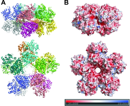
FIG. 4.
Overall view of the decameric arrangement of 1,3-PD dehydrogenase from K. pneumoniae. In both panels, the upper part corresponds to a side view, and the lower part shows a flat front view of the structure. (A) Ribbon representation of the decamer colored by monomer. (B) Electrostatic potential representation on the surface of the 1,3-PD dehydrogenase decamer, with the potential scale in the lower part of the panel (4). All figures were prepared using PyMol (12).

