FIG. 5.
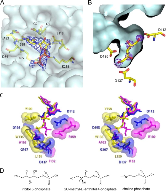
Ligand binding site of TarI. (A) The nucleotide pocket with bound CDP is shown with a composite-omit map rendered at 1σ, to show the presence of nucleotide. Residues involved in polar contacts to the nucleotide are shown in ball and stick representation. The surface of the protein is shown in light blue to highlight the pocket. (B) The ribitol pocket of TarI from the apo structure is shown with Fobs − Fcalc density for crystallographic waters, which correspond almost perfectly to potential sites of hydroxyl groups from a ribitol, which has been modeled by hand into the pocket. The corresponding contact residues are shown in stick representation with the ligand pocket surface shown in light blue. (C) Ligand binding pockets of cytidylyl transferases. Stereo overview of the ligand binding pockets is shown for those structures in the PDB with bound product. TarI (PDB 2VSH) is shown in blue, the MEP cytidylyl transferase IspD (PDB 1INI) is shown in magenta, and phosphorylcholine cytidylyl transferase LicC (PDB 1JYL) is in yellow. Key contact residues are labeled in the corresponding color for the protein. All ligands are shown in stick representation with contact residues shown in stick and sphere representation. (D) Structure of the substrates of cytidylyl transferases.

