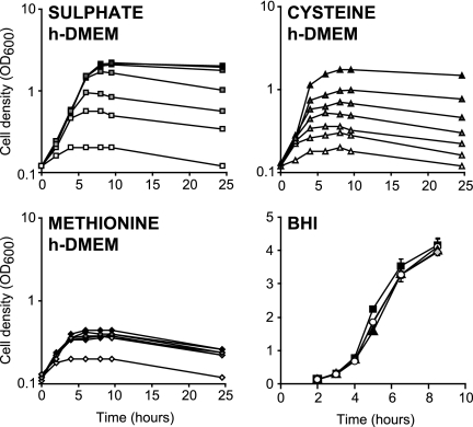
FIG. 3.
Effect of addition of different sulfur sources on growth of N. meningitidis B16B6. The growth of N. meningitidis B16B6 was followed by measuring optical densities in h-DMEM supplemented with different concentrations of K2SO4 (0, 25, 50, 100, 200, 400, and 800 μM from white to dark gray), methionine (0, 10, 20, 40, 80, 160, and 320 μM from white to dark gray), or cysteine (0, 10, 20, 40, 80, 160, and 320 μM from white to dark gray) and in BHI supplemented with a saturating concentration of methionine (20 μM) (▴), cysteine (200 μM) (⧫), or HCY (500 μM) (○), as well as in unsupplemented BHI (▪). Each growth curve is representative of three independent repeats.

