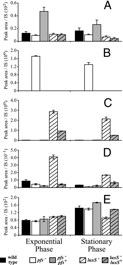
FIG. 4.
The profiles of AMC-related metabolites in pfs and luxS mutants of N. meningitidis are different. Cell extracts of the B16B6-pfs mutant (open bars), the B16B6-luxS mutant (open striped bars), wild-type strain B16B6 (black bars), the B16B6-pfs pfs+ complemented mutant (gray bars), and the B16B6-luxS luxS+ complemented mutant (gray striped bars) grown in BHI, prepared, and derivatized as described in Materials and Methods were analyzed by liquid chromatography-mass spectrometry to determine their SAM (A), SAH (B), SRH (C), HCY (D), and methionine (E) contents. The peak area corresponding to each compound in an extract was divided by the peak area of an appropriate internal standard (IS) for normalization; the data are the means ± standard errors for three independent cultures. Note that the complementing pfs gene in the B16B6-pfs pfs+ mutant is located closer to the origin of replication, which may have affected its expression level, and that expression of the complementing luxS gene in the B16B6-luxS luxS+ mutant is probably not driven by a complete set of the native luxS promoters. The scale on the y axis for in panel D precludes visualization of the ∼2-fold-lower concentration in the pfs mutant than in the wild type.

