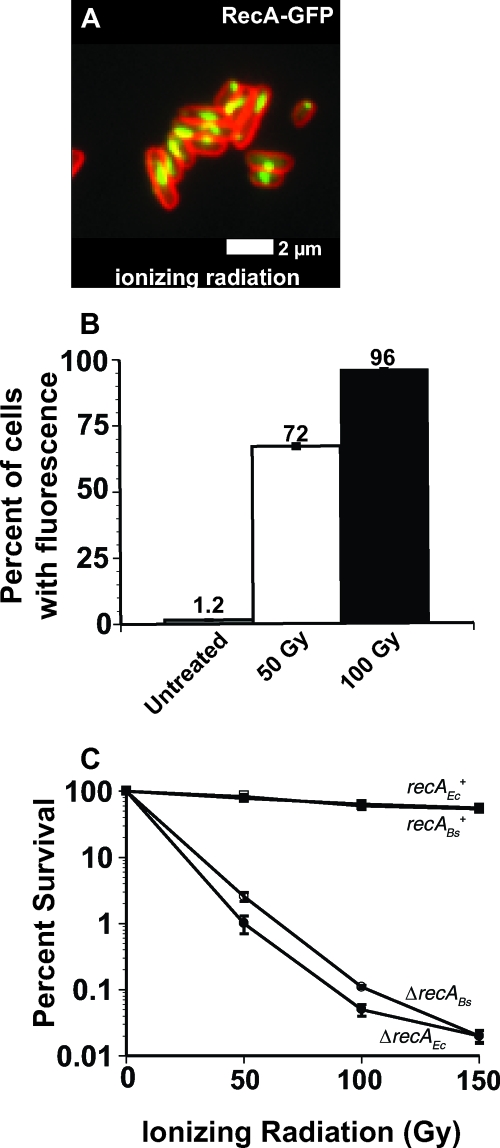
FIG. 1.
Ionizing radiation induces sulA promoter activity in E. coli. (A) Representative micrograph of E. coli RecA-GFP after ionizing radiation challenge. RecA-GFP is shown in green, and the cell membrane stained with FM4-64 is shown in red. White bar, 2 μm. (B) E. coli cells bearing the sulApΩgfp-mut2 promoter were used to monitor sulA promoter activity in response to DNA damage in single cells using fluorescence microscopy. The percentages of cells with sulApΩgfp-mut2 fluorescence are shown for untreated cells (n = 1,021) and cells after 50 Gy (n = 1,329) or 100 Gy (n = 1,420) of ionizing radiation. Error bars indicate ± the 95% confidence interval (variance) between at least three independent experiments as follows: untreated, 1.2 ± 0.14; 50 Gy, 72 ± 0.94; and 100 Gy, 96 ± 0.11. (C) Killing curve of wild-type E. coli (strain MC4100) and an isogenic strain with recA deleted. The open symbols represent B. subtilis recA+ (recABs+ [□]) and recA-deficient (ΔrecABs [○]) strains, and the closed symbols are E. coli recA+ (recAEc+ [▪]) and recA-deficient (ΔrecAEc [•]) strains. The error bars represent the standard errors from at least three independent experiments.

