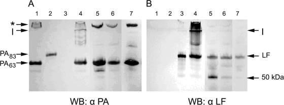
FIG. 1.
Detection of PA and LF in the plasma of a B. anthracis-infected monkeys, rabbits, and guinea pigs. Shown are results of SDS-PAGE followed by Western blot (WB) analysis of plasma from infected animals. Lane 1, PA63; lane 2, PA83, lane 3, LF; lane 4, in vitro-purified LT complex; lane 5, monkey plasma; lane 6, rabbit plasma; lane 7, guinea pig plasma. (A) Immunoblot stained with anti-PA monoclonal antibodies; (B) immunoblot stained with anti-LF monoclonal antibodies. An SDS-resistant oligomer of PA was detected in both the in vitro and in vivo LT complexes (marked by an asterisk). LF and a 50-kDa fragment of LF were also detected, most notably in plasma from the monkeys and rabbits. I, interface between the stacking and running gels; α, anti.

