
FIG. 4.
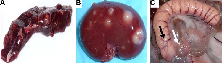
Gross pathology of extrapulmonary specimens on necropsy. (A) Spleen of a rabbit (AF4) infected with M. bovis AF2122. (B) Kidney of a rabbit (R1) infected with M. bovis Ravenel. (C) Appendiceal/cecal region of a rabbit (R3) infected with M. bovis Ravenel. The appendix (black arrow) and cecum (white arrow) are shown. Despite the cavity formation seen with other mycobacteria species and strains, only rabbits infected with M. bovis showed uniform spreading from primary lesions.