FIG. 1.
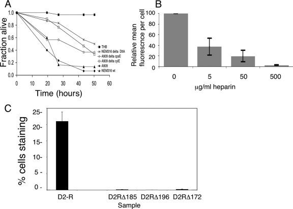
Drosophila as a model for GBS infection. (A) Male flies were infected with GBS and then incubated at 29°C. Data from a representative experiment with at least 30 flies per sample are shown. By Kaplan-Meier and log-rank analyses, median survival times for flies infected with wild-type GBS strains A909 and NEM316 were 21.5 and 27 h, respectively; strains with mutations in virulence determinants identified in mammalian models (cpsE, cylE, and dltA) were attenuated in Drosophila also, with median survival times of infected flies of 43.2, 43.2, and 80.2 h, respectively (P, <0.005 for each mutant versus the parent strain). wt, wild type. (B) Fluorescence-activated cell sorter analysis shows labeled-ACP binding to S2 cells with concentration-dependent inhibition by soluble heparin. Data are shown as the MFI ± SE per cell relative to that for untreated cells, normalized to the fluorescence of cells treated with labeled BSA. With 500 μg/ml heparin, the MFI was 2.5% ± 0.2% of that for untreated cells (P = 0.001; Student's t test). (C) Fluorescence-activated cell sorter analysis shows that the level of S2 cell binding to mutant D2-R constructs with charge-neutralizing mutations in GAG-binding residues (Arg185, Arg172, and Lys196 [D2RΔ185, D2RΔ172, and D2RΔ196]) was markedly lower than the level of S2 cell binding to the native D2-R protein. The data are expressed as the mean percentage ± SE of cells staining positive for the indicated protein.

