
FIG. 4.
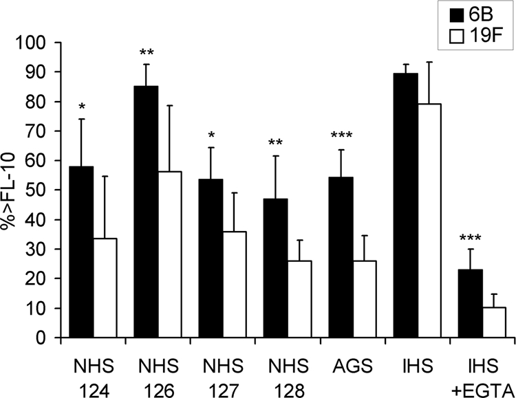
Comparison of C3 deposition on pneumococcal serotypes 6B and 19F with different sera. All serotype 6B and 19F strains (listed in Table 1) were analyzed in parallel with four different sera with low concentrations of anticapsular antibodies (NHS), an AGS, an IHS, and the IHS with 10 mM EGTA (IHS+EGTA). Average percentages of bacteria positive for C3 deposition (% > FL-10) with standard deviations are given. *, P < 0.05; **, P < 0.01; ***, P < 0.001 (Student's t test, 2-tailed with unequal variance).