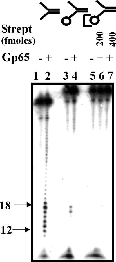
FIG. 3.
3′-5′ Exonuclease activity of gp65. Structure 1 (100 fmol), with either a free (lanes 1 and 2) or biotin-blocked 3′ arm (lanes 3 to 7) was treated with gp65 under standard conditions. The assay with the biotinylated substrate was performed either in the absence (lanes 3 and 4) or in the presence of the indicated amounts of streptavidin (lanes 6 and 7). Untreated and treated lanes are designated as “−” or “+”, respectively. In the structures at the top of the figure the circle represents a biotin moiety, and the third bracket represents biotin-bound streptavidin. The bands in the range of 12 to 18 nt arising out of junction cleavage are indicated on the left.

