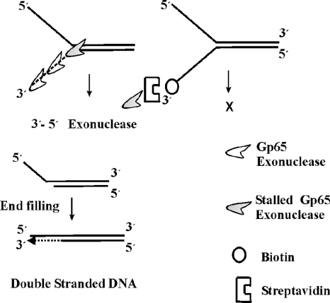
FIG. 8.
Proposed model depicting how gp65 processes fork junctions. gp65 is represented by an icon. The dashed line represents the exonucleolytically degraded 3′ arm. The protein travels in a 3′-5′ direction, serially cleaving the bottom strand until it reaches the junction, where it stalls (shaded icon). If the 3′ end of the fork is biotinylated and the biotin moiety is conjugated to streptavidin, the exonucleolytic activity of gp65 cannot function; no cleavage product is thus formed (figure on the right). The cleaved product is a structure in which there is a 5′ overhang. This can be potentially filled up by DNA polymerases.

