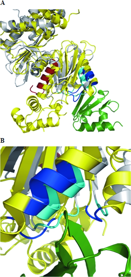
FIG. 3.
Superimposition of GlK on interaction between Mlc and PtsG. (A) Partial view of Mlc (yellow) in its interaction with the EIIB domain of PtsG (green) (31). The interacting amino acids between the two proteins are shown in blue. Glk (in gray) (26) has been superimposed onto the Mlc structure (48) by using the Coot program (http://www.ysbl.york.ac.uk/∼emsley/coot/). The amino acids contacting the EIIB domain are in cyan. The C-terminal helix harboring the 16 amino acids lacking in Glk15 (Δ305-321) are shown in red. The helix-turn-helix domain of Mlc (lacking in Glk) is on the lower left-hand corner. (B) Detailed view of the interaction site seen from the side opposite that shown in panel A.

