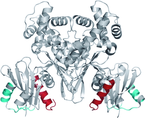
FIG. 5.
Structure of Glk. The structure of the Glk dimer (gray) (26) is shown. The C-terminal helix harboring the 16 amino acids lacking in Glk15 (Δ305-321) are shown in red. They most likely constitute the interaction domain with MalT. The putative interaction site with EIIB of PtsG is indicated in cyan.

