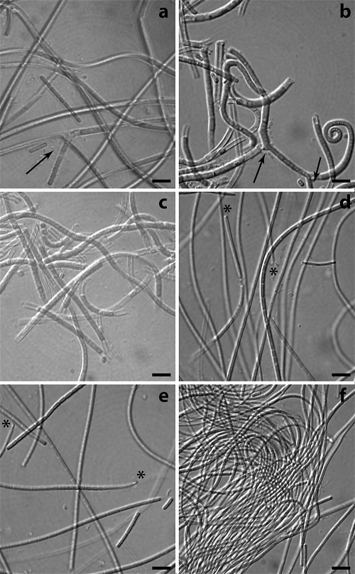
FIG. 1.
Photomicrographs illustrating the morphological features of the six Leptolyngbya strains. On the basis of their pigmentation they could be divided into phycocyanin-rich strains (green) VRUC184 (a), VRUC201 (b), and VRUC206 (c) and phycoerythrin-rich strains (red) VRUC192 (d), VRUC198 (e), and VRUC135 (f). Arrows indicate false branching (a and b), and asterisks indicate the eyespot-like structure at the tip of the apical cell (d to f). Bars, 5 μm.

