Figure 2.
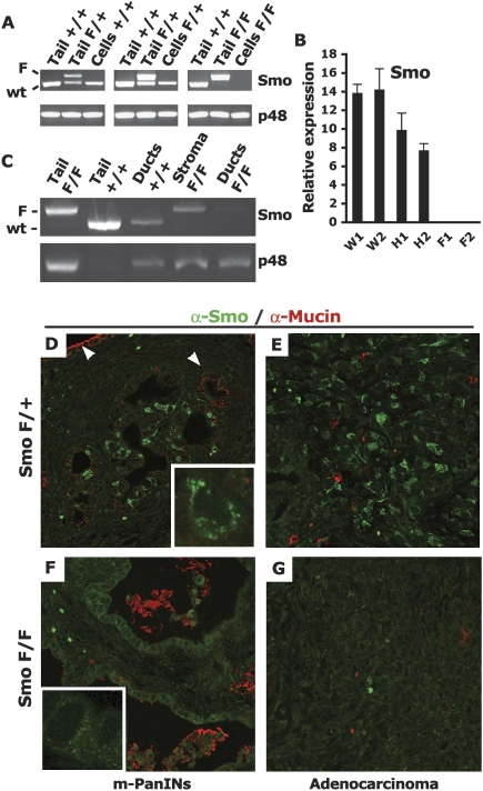
Smoothened is depleted in ductal cells of PDAC SmoF/F mice. (A) Recombination of the Smo locus in PDAC cell lines. PCR amplification of the Smo locus (Smo) or the p48-Cre transgene (p48). The Smo genotyping procedure amplifies the nonrecombined conditional SmoF allele (upper band, F) and the wild-type (wt) Smo allele (lower band, wild type). The upper PCR band is lost upon Cre recombination of the Smo conditional locus. The input genomic DNA used in each PCR reaction is indicated: (Tail) genomic DNA from mouse tail, 100 ng; (Cells) genomic DNA from PDAC-derived tumor cell lines, 10 ng; (+/+) Smo+/+; (F/+) SmoF/+; (F/F) SmoF/F. (B) Depletion of the Smo mRNA in recombined cell lines. Expression of Smo mRNA in total RNA extracts from Smo+/+ (W), SmoF/+ (H), or SmoF/F (F) PDAC cell lines. Levels of mRNAs expressed as a percentage of the m-Gus control mRNA. Total RNA extracts from two cells lines of each genotype were assayed in triplicate. (C) In vivo recombination of the Smo locus. Genomic DNA from ductal structures or stromal areas isolated by laser-capture microdissection (LCM) from PDAC tumors was subjected to the same PCR amplification as in A. Ducts or stromal areas from two PDAC tumors of each genotype were pooled and subjected to PCR amplification. The input genomic DNA used in each PCR reaction is indicated: (Tail) DNA from mouse tail; (Ducts) LCM-captured ducts from two PDAC tumors; (Stroma) LCM-captured stromal-rich area of two PDAC tumors; (+/+) Smo+/+; (F/+) SmoF/+; (F/F) SmoF/F. (D–G) In vivo depletion of the Smo protein. Immunofluorescent detection of Smo (green) and Muc-1 (red) (630×). The Smo protein is detected in a subset of mucin-negative ducts inside PDAC SmoF/+ PanIN-like lesions (D), but not in mucin-positive ducts (white arrows) as well as in mucin-negative PDAC SmoF/+ adenocarcinoma lesions (E). Smo is undetectable in PDAC SmoF/F PanIN-like lesions (F) orin PDAC SmoF/F adenocarcinoma (G). Granular cytoplasmic Smo staining of an individual PDAC SmoF/+ cell (2520×) (D, insert) absent in individual PDAC SmoF/F cells (F, insert). The panels are representative of multiple fields of pancreatic sections from three PDAC SmoF/+ mice and three PDAC SmoF/F mice.

