Figure 3.
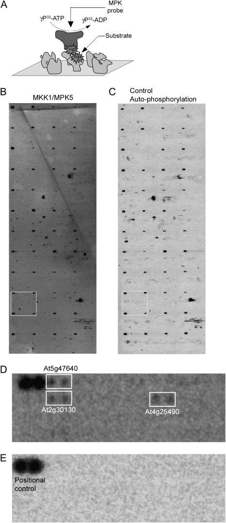
Identification of MPK phosphorylation substrates on protein microarrays. (A) Schematic of kinase assays on protein microarrays. MPK-TAP fusion proteins were purified from N. benthamiana overexpressing 20 selected in vitro functional combinations of MKK/MPK. Purified MPK proteins in kinase buffer were overlaid on protein microarrays in the presence of [γ-33P]-ATP and incubated for 1 h at 30°C. Phosphorylated MPK substrates on the protein microarrays were detected by exposure to Kodak film. (B,C) A representative microarray probed with activated MPK5 (B) and a representative negative control overlaid with kinase buffer and [γ-33P]-ATP to detect autophosphorylating proteins (C) are shown. Bright dark spots at the corner of each block represents positional controls spotted in duplicate to help with subsequent grid alignment and spot identification. (D,E) Zoomed-in area of block 26 marked as white rectangle from microarrays represented in B and C are shown in D and E, respectively. White rectangle regions in D represent putative MPK5 phosphorylation targets for which signal is present in the MPK5 probed array (D) but absent in the autophosphorylation control array (E).

