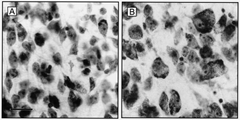
Figure 2.
Representative photomicrographs of cresyl violet-stained sections of the infundibular nucleus of young, premenopausal (A) and older, postmenopausal (B) women. Note the considerably enlarged neurons in the older subject with increased size of nuclei and nucleoli as well as increased Nissl substance. Scale bar = 25 microns in A (applies to A,B). From Abel and Rance [2], reproduced with permission from Wiley-Liss Inc.

