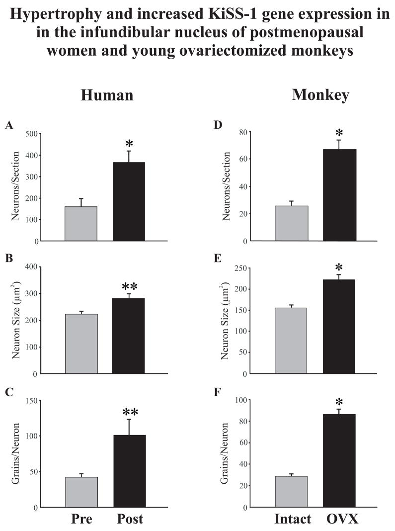
Figure 6.
Changes in neuronal morphology and KiSS-1 gene expression in the infundibular nucleus of premenopausal and postmenopausal women (A,B,C) or the infundibular nucleus of young, intact and ovariectomized cynomolgus monkeys (D, E, F). Figure 6A shows the mean number of neurons expressing KiSS-1 mRNA in sagittal human sections and 6D shows the mean number of neurons in unilateral coronal sections in the monkey. 6B and E show the mean profile area (μm2) of KiSS-1 neurons and C and F show the mean number of autoradiographic grains for each labeled neuron. Postmenopausal women exhibited increased number, size and gene expression of KiSS-1 neurons that was similar to that seen in young, ovariectomized cynomolgus monkeys. Values are expressed as mean ± SEM. * Significantly different from premenopausal (A) or intact (D, E, F), p< 0.001. ** Significantly different from premenopausal (B, C), p< 0.05.
From Rometo et al. [86], reproduced with permission from The Endocrine Society.

