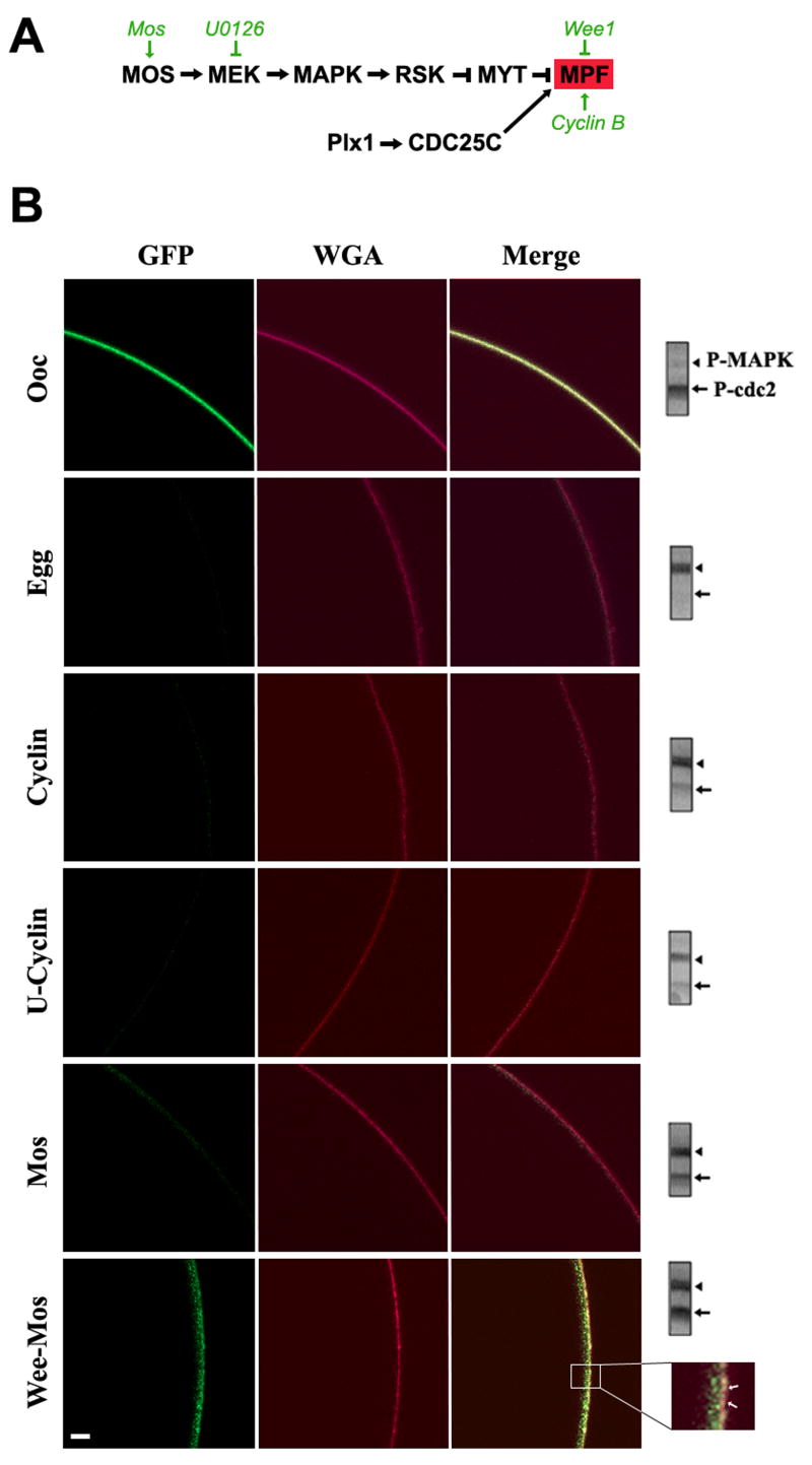
Figure 3. Kinase-dependent modulation on PMCA internalization.
A. Signaling cascade regulating Xenopus oocyte meiosis entry. The steps at which the molecular and pharmacological manipulations used act are shown in green. B. Live cell imaging of PMCA internalization. Oocytes expressing GFP-PMCA were subjected to one of the following treatments: injected with Cyclin B1 RNA (Cyclin); pre-treated with U0126 (50μM) for 1hr before cyclin RNA injection (U-Cyclin); injected with Mos RNA (Mos); or injected with Wee1 RNA and incubated overnight before Mos RNA injection (Wee-Mos). The cell membrane was stained with WGA-Alexa633. The inset shows that in the Wee-Mos treatment, GFP-PMCA internalization is delayed but not completely inhibited. The examples shown are representative of 76 and 29 oocytes and eggs respectively, and 11-15 cells each for treatments used to manipulate the kinase cascades. All cells analyzed showed a similar pattern of PMCA localization to the examples shown, except for the Wee-Mos treatment where in 3/11 cells PMCA was internalized to similar levels as Mos. Western blot analysis for phospho-MAPK (P-MAPK) and phospho-MPF (P-MPF) of each of the scanned cells is shown on the right. The equivalent of ½ a cell was loaded. The scale bar is 20μm.

