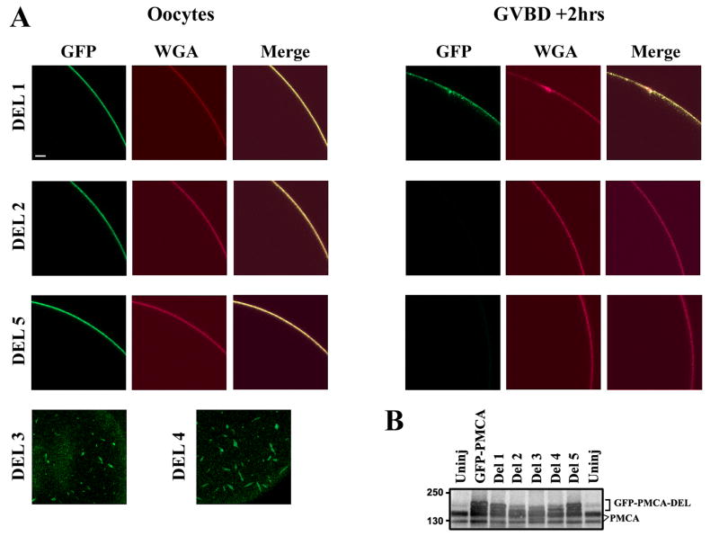
Figure 5. Deletion Analyses.
A. Oocytes were injected with RNA (5-7ng) of each of the GFP-PMCA deletions (DEL1, DEL2, and DEL5), then activated with progesterone 4-5days after injection. Internalization was assayed by imaging cells stained with WGA-Alexa633. For DEL3 and DEL4 no signal was observed at the cell membrane following injection of different RNA concentrations up to 12 days for DEL3 and 16 days for DEL4. Rather the products of these deletions localize intracellularly to elongated vesicle-like structures (Del3, Del4). The examples shown are representative of 5-10 cells for each deletion. Scale bar is 20μm. B. Expression of the full length and deletions. Anti-PMCA blot from uninjected oocytes (Uninj), oocytes injected with full-length GFP-PMCA and the 5 deletions. For the full-length, DEL1, DEL2, and DEL5 lysates were collected 5-7 days after injection. DEL3 and DEL4 lysates were collected 9 days and 16 days post-injection respectively. The bracket marks the electrophoretic mobility of full-length and different deletion mutants (GFP-PMCA-DEL). Endogenous PMCA is also indicated (PMCA).

