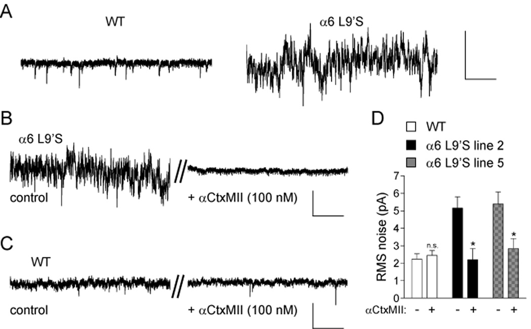
Figure 5. Spontaneous α6 channel activity in α6L9’S VTA DA neurons.
(A) Increased current fluctuations in voltage-clamp recordings from α6L9’S neurons. VTA DA neurons from WT or α6L9’S mice were voltage clamped at −60 mV. A segment from a representative voltage clamp recording is shown. Scale bars: 40 pA, 0.8 s.
(B) Noise in α6L9’S neurons is dependent on α6* nAChRs. VTA DA neurons were voltage clamped at −60 mV and a baseline recording was taken (control). αCtxMII was bath applied for 10 min, and a representative (n = 4, each line) segment of the voltage clamp record after this 10 min period is shown (+αCtxMII, 100 nM) for the same cell.
(C) αCtxMII inhibition has no effect in WT control recordings. As a negative control for perfusion artifacts, WT neurons were assayed as in (B).
(D) Quantification of channel noise increase in α6L9’S mice. RMS noise values for voltage clamp recordings from VTA DA neurons in the presence and absence of αCtxMII is shown. Data are reported as mean ± SEM. *, p < 0.05. Scale bars (B and C): 20 pA, 0.5 s.

