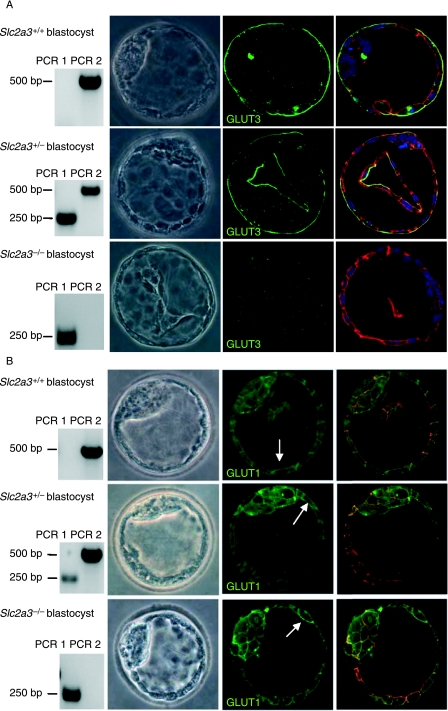
Figure 3.
Detection of GLUT3 and GLUT1 in Slc2a3+/+, Slc2a3+/−, and Slc2a3−/− blastocysts at day 4·5 pc. Blastocysts were isolated and stained for (A) GLUT3 or (B) GLUT1 in combination with an Alexa 488-labeled secondary antibody. Genotyping (left panels) was performed by PCR as described in the legend of Fig. 2. Nuclei (blue) and actin cytoskeleton (red) were counterstained. GLUT1 immunoreactivity is indicated by arrows.

