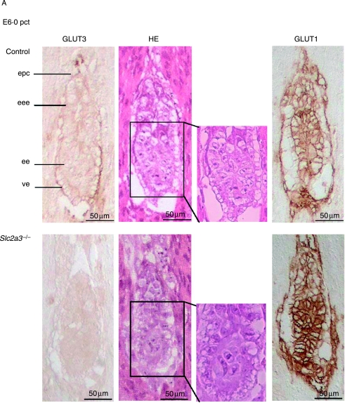
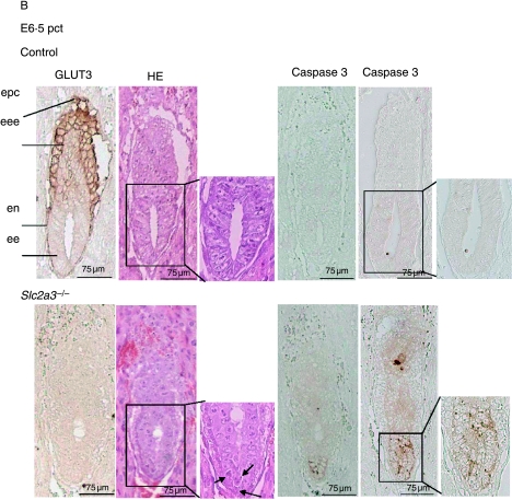
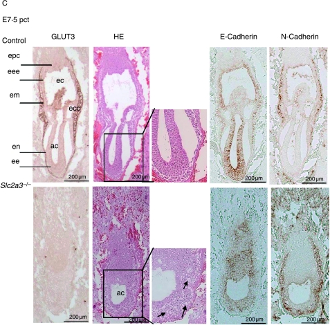
Figure 4.
Defective development and apoptotic cell death in Slc2a3 mutant embryos. Histological analysis of Slc2a3−/− and control embryos (either Slc2a3+/+ or Slc2a3+/−). Serial sagittal sections of uteri from heterozygous matings at (A) day 6·0, (B) 6·5, and (C) 7·5 pc were stained with hematoxylin and eosin (HE), or used for staining of GLUT3, GLUT1, activated caspase 3, E-cadherin, or N-cadherin. ac, amniotic cavity; ec, ectoplacental cavity; ecc, exocoelomic cavity; ee, embryonic ectoderm; eee extraembryonic ectoderm; em, embryonic mesoderm; en, endoderm; epc, ectoplacental cone. Microscopical abnormalities in ectodermal cells of mutant embryos are indicated by arrowheads.


