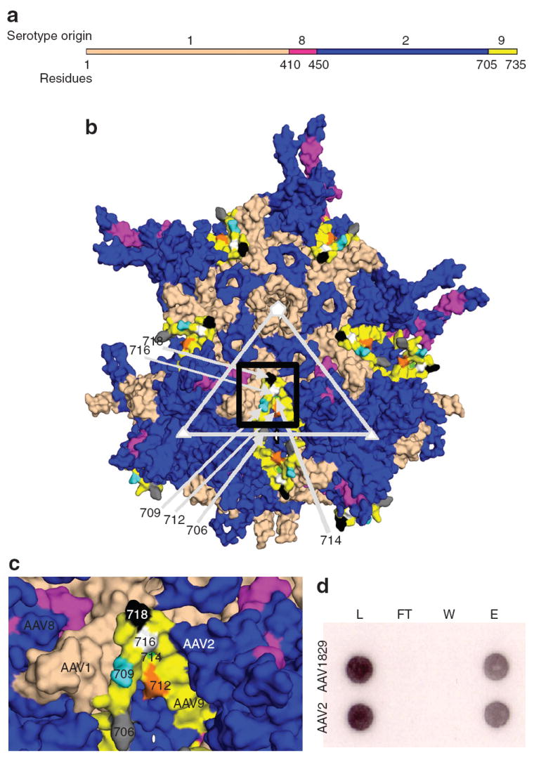
Figure 2. Physical characterization of chimeric-1829.

(a) Primary structure of novel adeno-associated virus (AAV) variant selected from CS1cell. (b) Three-dimensional model showing VP3 subunits of AAV1829 in relation to the fivefold (white pentagon); threefold (white triangle) and twofold (white oval) axes of symmetry. AAV1-derived residues are colored salmon, AAV2 in blue, AAV8 in dark pink, and AAV9 in yellow. (c) Black box region in 2. Panel b shown at higher magnification. Key residues in the C-terminal AAV9 region that differ from AAV2 are colored gray (706); cyan (709); orange (712); green (714); white (716); and black (718) using Vp1 numbering. (d) Heparin binding profiles of AAV1829 and AAV2. L, load; FT, flow through; W, wash; e, elution.
