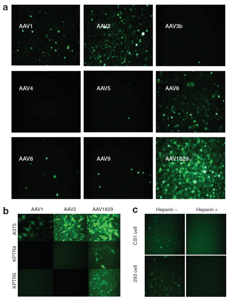
Figure 3. Biological characterization of chimeric-1829 compared with some parental serotypes.

(a) Fluorescence micrographs of green fluorescent protein (GFP) transgene expression in CS1 cells transduced with AAV serotypes 1–9 (except 7) and the novel variant chimeric-1829 at a multiplicity of infection (MOI) of 1,000 for 48 hours. (b) Representative fluorescence micrographs of GFP transgene expression in different human and mouse melanoma cell lines infected with chimeric-1829 and parental serotypes at an MOI of 1,000 for 48 hours. (c) Heparin-inhibition assay. Top panels show CS1 cells, and bottom panels show 293 cells. Left and right columns are transduction profiles in the absence and presence of heparin, respectively.
