
Figure 1.
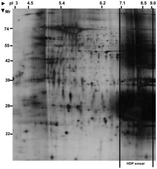
2D gel image of total P. falciparum lysate run on a wide range pI3-10 immobiline dry strip for first dimensional focusing. The approximate molecular weights and pI values were derived from protein identifications of the gel spots. Significant smearing resulting from the hemeoglobin-derived products (HDP) is seen in the basic region of the gel (pI 7.0-9.0). The high abundance of these products, while masking the basic plasmodial proteome, also limits protein loading capacities, resulting in poor spot intensity and resolution in other regions of the gel.