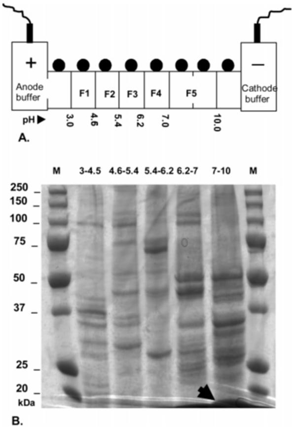
Figure 2.

Use of solution isoelectric focusing for HDP containment. (A) Schematic representation of the ZOOM-IEF focusing device showing the 5 chamber arrangement used in this study. The high protein concentration due to compartmentalization of the high abundance HDP to fraction 5 (pI 7-10) required doubling of the volume of this chamber using a spacer. The pH values refer to the type of the membrane disk used at this point. First and last chambers were not loaded as per manufacturer’s instructions. (B) 1D gel loading of proteins precipitated from 50 μL of each of the five post-fractionation samples (F1: 3.0-4.5, F2: 4.6-5.4, F3: 5.4-6.2, F4: 6.2-7.0, F5: 7-10). The differential banding observed indicates efficient fractionation. The arrow shows the position and compartmentalization of HDP with unmasking of other proteins in the fraction. Lanes M: markers with molecular weights as indicated.
