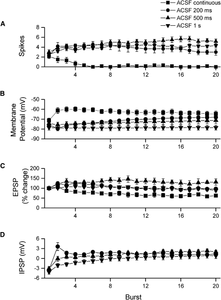
Figure 7.
Summary of the postsynaptic effects of continuous tetanization and burst stimulation in standard ACSF. For analysis of cells receiving continuous tetanization, the 80 stimuli were divided into 20 segments. These 20 segments correspond to the discrete bursts given during burst stimulation. (A) All stimulation conditions evoked an equivalent number of spikes on the first burst; however, spike firing during continuous tetanization declined with repeated stimulation, and very little firing was seen after the first four bursts. For all other stimulation conditions, firing increased across the first several bursts and then remained relatively constant through the final burst. (B) Membrane potential measured immediately before each burst showed substantial differences depending on stimulation condition. With continuous 100-Hz tetanization, fast, AMPA receptor-mediated EPSPs summed across all 80 stimuli to produce a large depolarization. This depolarization reached a peak over bursts 2–5. However, as EPSPs began to depress (C), membrane potential gradually declined back toward the resting level. For burst stimulation at 200-msec intervals, the second burst was given before the IPSP from the first burst had fully decayed and so the response to the second burst began from a hyperpolarized membrane potential. However, as bursts were repeated, membrane depolarization summed across the remaining 18 bursts. Burst stimulation at 500-msec intervals had a similar, though smaller, effect on membrane potential. The second burst began at a slightly hyperpolarized membrane potential, because the IPSP following the first burst had almost, but not completely, decayed. The wider spacing of bursts also resulted in less efficient summation across bursts, although membrane potential eventually became depolarized over bursts 10–20. Burst stimulation at 1-sec intervals caused very little change in membrane potential because individual bursts were too widely spaced to be affected by preceding IPSPs or EPSPs. (C) Continuous 100-Hz tetanization caused a gradual depression of EPSP slopes. Depression was observed as early as the third burst and continued to develop until around burst 10–12. In contrast, burst stimulation at 200- and 500-msec intervals initially facilitated EPSPs (bursts 2–8); however, facilitation faded for cells receiving burst stimulation at 200-msec intervals but remained for cells receiving burst stimulation at 500-msec intervals. Cells receiving burst stimulation at 1-sec intervals showed little change in EPSPs across bursts. (D) During burst stimulation there was a gradual, cumulative decrease in post-burst inhibition. The rate of decrease was fastest for the 200-msec burst interval, where post-burst inhibition was completely suppressed by the second burst, somewhat slower for the 500-msec burst interval, and slowest for the 1-sec burst interval.

