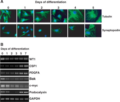
Figure 2.
Differentiation of MPC5 cells. (A) Mouse MPC5 cells were differentiated for the times indicated at top. The cells were then fixed and subject to immunofluorescence with either anti-tubulin or anti-synaptopodin antibodies (both in green), counterstaining with Hoechst. (B) MPC5 cells were differentiated for the number of days indicated and total RNA prepared. Semi-quantitative RT-PCR was performed to detect WT1, CSF1, PDGFA, Bak, c-myc, Podocalyxin and GAPDH.

