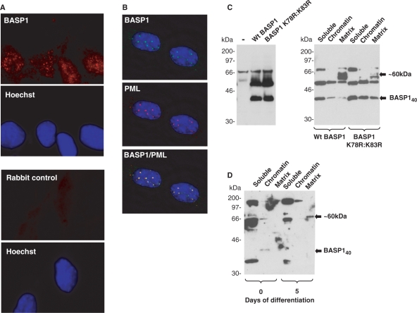
Figure 6.
Sumoylation of BASP1 affects its intranuclear localization. (A) Immunofluorescence was performed with MPC5 cells using rabbit anti-BASP1 antibodies (top, red) or secondary antibody alone (bottom). Hoechst staining for each panel is shown below. (B) MPC5 cells were subject to immunofluorescence with sheep anti-BASP1 (green) and rabbit anti-PML (red) antibodies. A merge of the two images is shown in the bottom panel. The images were captured using a Deltavision microscope. (C) Wild-type HA-tagged BASP1 or the mutant derivative (K78R:K83R) were transfected into K562 cells. Forty-eight hours later either whole-cell extracts (left) or nuclei were extracted and subfractionated (right). Samples were resolved by electrophoresis and immunoblotted with anti-BASP1 antibodies. Molecular weight markers (in kilodalton) are shown at left. BASP140 and BASP1 ∼ 60 kDa are indicated by arrows. (D) Undifferentiated MPC5 cells or MPC5 cells that had differentiated for 5 days were analysed as in (C) to detect endogenous BASP1 in the soluble, chromatin and matrix compartments of the nucleus. BASP140 and the ∼60-kDa form of BASP1 are indicated.

