Figure 2.
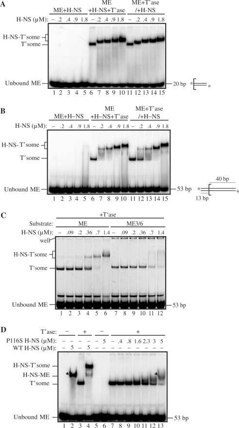
Electrophoretic mobility-shift assays with Tn5 transpososomes and H-NS. (A) and (B) H-NS mobility-shifts of transpososomes formed with 20 and 53 bp ME substrates, respectively. Where indicated, H-NS was added to transpososome assembly reactions either at the same time as transposase (ME+T’ase+H-NS) or after transposase (i.e. subsequent to transpososome formation) (ME+T’ase/+H-NS). An illustration of the respective substrates is shown beside the gel image; the bracket defines the beginning of the ME sequence and asterisks indicate the position of 32P-labels. (H-NS-T'some) H-NS bound transpososome; (T'some) transpososome. (C) H-NS mobility-shift of a transpososome formed with a ME substrate containing a G to C and a T to A mutation at positions 3 and 6, respectively (ME3/6). (D) H-NS mobility-shift with P116S H-NS. The arrowheads show the mobility of a non-specific H-NS-ME complex (lane 2) and a P116S H-NS-transpososome complex (lane13). H-NS binds the ME in the absence of transposase only at relatively high (⩾5 μM) H-NS concentrations. Similarly, P116S H-NS binds to the transpososome only at relatively high concentrations (⩾3 μM). Note that in (A)–(D) HA transposase was used.

