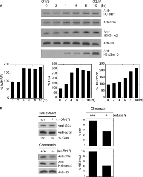
Figure 3.
UHRF1 affects chromatin association of G9a. (A) Concurrent loading of hUHRF1 and G9a onto chromatin during S phase. HeLa cells were synchronized by double aphidicolin/thymidine block and released to regular medium for hours indicated at the top of the panel. The chromatin fractions at each time point were used for western-blot analysis with antibodies indicated. Anti-H3 and H3 phopho-Ser10 (H3 pSer10) antibodies were used for a loading control and mitosis marker, respectively. Densitometric scans of hUHRF1, G9a and H3K9me2 levels in each chromatin fraction are shown after normalization by the corresponding H3 level. (B) Impaired chromatin association of G9a in the absence of mUHRF1. Cell extracts and chromatin were prepared from wild type (+/+) and mUhrf1-null (−/−) ES (ES) cells and used to detect the presence of G9a and dimethylated H3K9 (H3K9me2) with antibodies indicated. The relative G9a level in cell extracts is shown as percentage G9a at the bottom of the panel by a densitometric analysis after normalization by the actin-loading control. Normalized densitometric scans of G9a and H3K9me2 levels in chromatin are shown in graphs.

