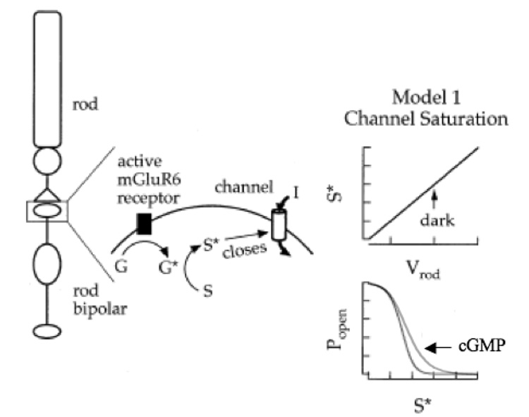
Fig. 3. Model depicting the proposed role of cGMP in mGluR6 transduction.
Left: Binding of glutamate, the rod transmitter, to mGluR6 activates Go (G*), which in turn catalyzes the production of a substance (S*) that closes the synaptic channel. Right: In this model, there is a linear relationship between the rod membrane potential (which controls transmitter release) and the production of S* (top). At depolarized rod membrane potentials (i.e., in the dark) the production of S* is in excess compared with the number of channels, allowing for negative-going excursions of the rod membrane potential without significant channel opens (bottom). cGMP/cGK functionally shifts this curve to the right, allowing for small changes in membrane potential to open channels, but does not change the maximum open probability. Modified from (Sampath and Rieke, 2004).

