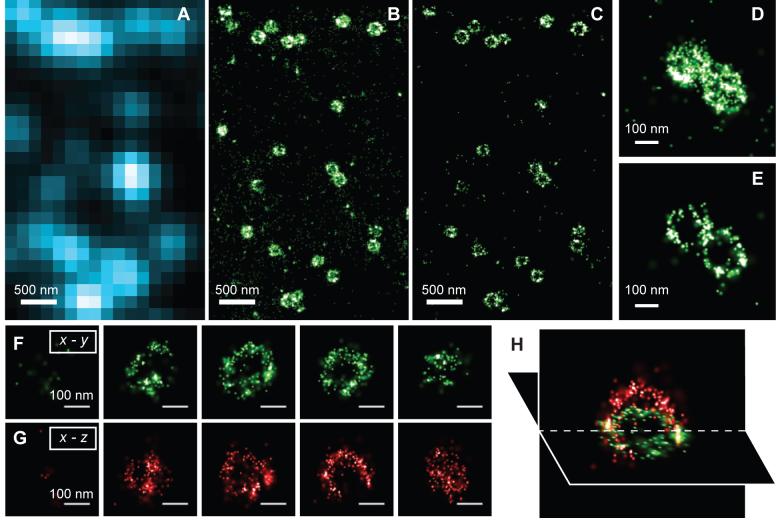
Fig. 3.
Three-dimensional STORM imaging of clathrin-coated pits in a cell. (A) Conventional direct immunofluorescence image of clathrin in a region of a BS-C-1 cell. (B) The 2D STORM image of the same area with all localizations at different z positions included. (C) A x-y cross-section (50 nm thick in z) of the same area showing the ring-like structure of the periphery of the CCPs at the plasma membrane. (D, E) Magnified view of two nearby CCPs in 2D STORM (D) and their 100 nm thick x-y cross-section in the 3D image (E). (F - H) Serial x-y cross-sections (each 50 nm thick in z) (F) and x-z cross-sections (each 50 nm thick in y) (G) of a CCP, and an x-y and x-z cross section presented in 3D perspective (H), showing the cage like structure of the pit.

