
Figure 3. Tumorigenesis in the mouse small intestine is initiated in Prom1+ crypt stem cells.
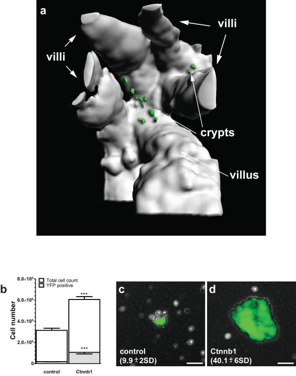
(a) View (394μm depth) into the base of a small intestinal crypt in a Prom1+/C-L ; RosaYFP ; Ctnnb1+/lox(ex3) mouse, two days following tamoxifen-induction. Mice had 8.4 YFP+ cells per crypt (range 2 to 16 >210 crypts counted). (b) Graph reporting the mean (±s.e.m., n=3) number of total and YFP+ cells isolated 2 days following tamoxifen induction, from Prom1+/C-L ; RosaYFP ; Ctnnb1+/+ (control) and Prom1+/C-L ; RosaYFP ; Ctnnb1+/lox(ex3) (Ctnnb1) small intestinal mucosae (***=P<0.0001, Wilcoxon test). Colonies formed following four weeks in stem cell culture medium by single cells isolated 2 days post tamoxifen induction from Prom1+/C-L ; RosaYFP ; Ctnnb1+/+ (c) and Prom1+/C-L ; RosaYFP ; Ctnnb1+/lox(ex3) (d) small intestinal mucosae, numbers=total colonies per small intestinal culture (±s.e.m., n=10, P<0.0001, Wilcoxon test). Scale bars=50μm.