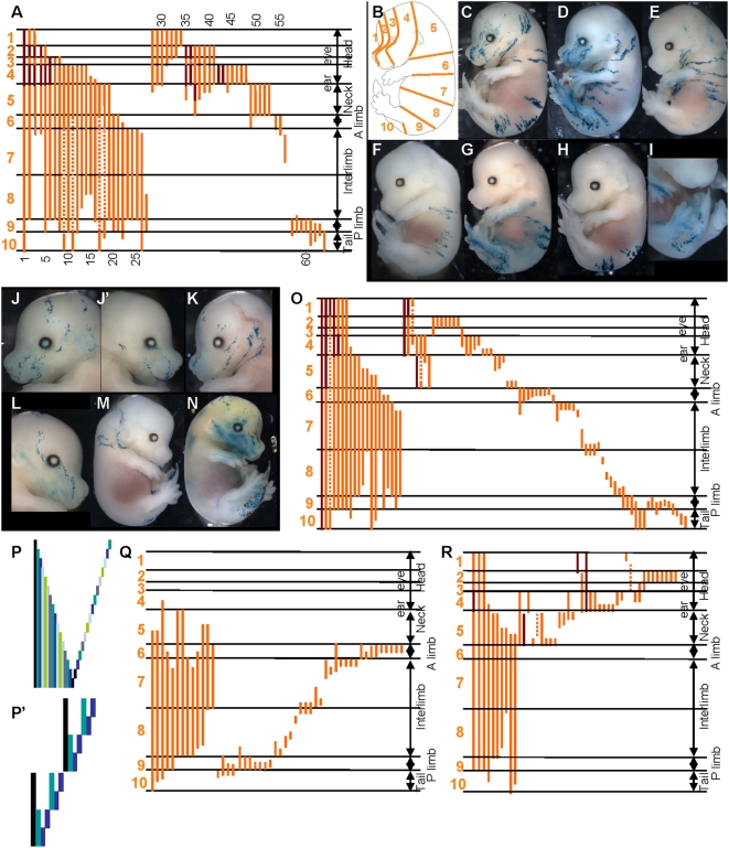
Figure 3. Three pools of SE-forming cells at E6-5-E7.5, following distinct modes of growth in the head and the trunk.
(A) Schematic representation of the pattern of the 64 clones in E14.5 lox-LacZ embryos induced at E6.5–E7.5. Horizontal lines represent the boundaries between the regions of the body in A. Each vertical orange line corresponds to a single clone; no contribution to the levels where the line is interrupted. Dark red lines represent the contribution of the clone to the contralateral side. Clones were first classified according to size (long on left and short on right) and then according to the most anterior region to which they contribute. (B) Schematic representation of an E14.5 embryo showing the regions used in A. (C)–(M) Examples of E14.5 lox-LacZ embryos. (C)–(G) and (M) long clones; (H) and (I) posterior short clones; (J)–(L) anterior short clones. (N) is a spontaneous clone (in a CT2 embryo) labelled only in the head and the tail. (O) Schematic representation, as in fig. 4, of the pattern of the clones in E14.5 LaacZembryos. All clones were classified according to size (long on left and short on right) and then according to the most anterior region to which they contribute. (P), (P′) Pattern of clones expected from the labelling of cells in the pool of precursors classified according to the most posterior region to which they contribute for two representative models for their production: self-renewing pool of cells (P); and from the regional mode (P′). Each column (of different colour) represents a clone. (Q)–(R) Schematic representation, of the LaacZ clones that contribute (Q) to regions 6 to 9 (see B); (R) to regions 1 to 5 (see B). Clones were classified according to size (long on left and short on right) and the most posterior region to which they contribute. Note that clones from ancestral cells of the SE (shown in O) have been removed. (Q) Long clones that contribute to all regions are present on the left. (R) Clones from ancestral cells of head SE are shown on the left. Note the absence of clones that contribute to all five sub-regions.

