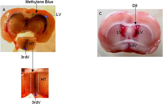
Figure 3. Representative images of brains sectioned after lateral ventricle administration with either methylene blue or DiI dyes.
A, Coronal section of adult rat brain injected icv with methylene blue, showing the dissected hypothalamus (HT), (LV: lateral ventricle), (3rdV: third ventricle). B, Enlargement of HT showing the region (dotted lines) that was removed prior to starting cell culture. C, Coronal section of adult rat brain after icv DiI injection, laterals and third ventricles are red stained by the dye.

