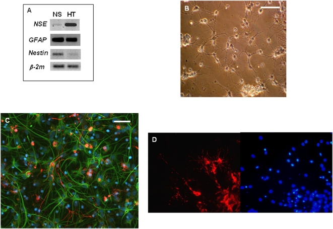
Figure 4. Hypothalamic NS cultures harbor precursors that can differentiate in neurons and glial cells.
A: Gene expression analysis of some lineage-specific CNS markers by RT-PCR. RNA was isolated from hypothalamic-derived fetal secondary NS and adult hypothalami (HT) used for comparison: NSE, a marker for neurons, GFAP, a marker for astrocytes and Nestin, a marker for undifferentiated/neuroepithelial cells. β-2 microblobulin ( β-2m) was used as housekeeping gene. B: Phase-contrast image showing the typical morphology of differentiating NS cultures after 7DIV. C: Representative fluorescence micrograph after double-labeling immunofluorescent detection of neuronal and astrocyte cells in fetal differentiating cultures (7DIV) by using antibody lineage-specific markers: βTubIII (red: neurons) and GFAP (green: astrocytes). Nuclei are stained in blue by the dye DAPI. D, fluorescence micrograph after immunofluorescent detection of oligodendrocyte cells (showned here in red) in fetal differentiating cultures (7DIV) by using an antibody oligodendrocyte-specific marker: O4 (left side). DAPI-stained nuclei from same micrograph shown on right side. Scale bars: 50 μm.

