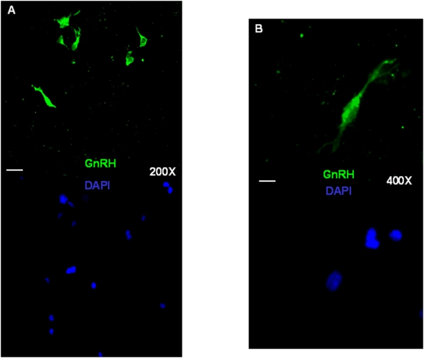
Figure 6. GnRH-immunoreactive cells as detected by a specific anti-GnRH antibody in differentiating fetal hypothalamic NS cultures.
Representative fluorescence micrographs showing some GnRH-labeled immunofluorescent cells (green fluorescence) as detected in differentiating cultures kept for 5 days in DMEM/F12, FGF-2 (20ng/mL) followed by 5 days in DMEM/F12, B27 (0.5%). Duplicate pictures show the nuclei stained in blue by the dye DAPI. Scale bars: 20 μm (A panel) and 10 μm (B panel).

