Figure 5.
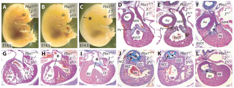
Both Pbx2 and Pbx3 contribute to OFT septation and semilunar valve formation. A though C, Gross appearance of Pbx2−/−3+/− (A), Pbx1+/−2−/− (B), and Pbx1+/−2−/−3+/− (C) embryos at E14.5. Asterisk indicates generalized edema. D through F, Transverse sections of the RVOT at E14.5. Pbx2−/−3+/− (D) and Pbx1+/−2−/− (E) embryos display patent RVOTs with slender pulmonic valve (PV) leaflets. The RVOT of Pbx1+/−2−/−3+/− embryos (F) is narrowed and associated with malformed pulmonic valve leaflets. G though I, Transverse sections of the right (RV) and left (LV) ventricles at E14.5. Both Pbx2−/−3+/− (G) and Pbx1+/−2−/− (H) mice have normal right and left ventricular walls. Pbx1+/−2−/− mice (H) exhibit a ventricular septal defect (asterisk). Pbx1+/−2−/−3+/− embryos (i) have both a ventricular septal defect (asterisk) and right ventricular hypertrophy. J through L, Transverse sections of the aortic valve at E14.5. Pbx2−/−3+/− mice (J) have a normal trileaflet aortic valve. Pbx1+/−2−/− mice (K) have a bicuspid aortic valve. Pbx1+/−2−/−3+/− embryos (L) have malformed aortic valves (AV). NCC indicates noncoronary cusp; RCC, right coronary cusp; LCC, left coronary cusp.

